Open trap of aquatic carnivorous plant, humped bladderwort Utricularia gibba, with single-cell organisms inside. Technique: Confocal imaging, 100x. Shot by Dr. Igor Siwanowicz.While the above image may seem like some sort of amazing blacklight poster from the '70s, in actuallity it's a close up shot of a vicious, carnivorous plant. A "Humped Bladderwort" to be specific--and the winner of the first prize in 2013's Olympus BioScapes Digital Imaging Competition. An annual contest that challenges both scientists and every day nature enthusiasts to find beauty in the tiniest of microbes, the Humped Bladderwort beat out over 2,100 still images submitted from 71 countries for the grand prize and unofficial title of "the hottest thing under a microscope."
Advertisement
Captured by Igor Siwanowicz, a scientist at the Howard Hughes Medical Institute's Janelia Farm Research Campus, the shot looks almost like a CGI generated picture or fractal image.Proving that sometimes nature can be more beautiful than anything man can imagine (zebras anyone?) check out some of the other shots that made the final cut below:
Single-cell fresh water algae (desmids). Composite image including, concentric from the outside: Micrasterias rotata, Micrasterias sp., M. furcata, M. americana, 2x M. truncata, Euastrum sp. and Cosmarium sp. Technique: Confocal imaging, 400x. Shot by Dr. Igor Siwanowicz.
Lily flower bud, transverse section. Technique: Darkfield illumination, stitched images, shot by Mr. Spike Walker.
Mouse embryonic fibroblasts showing actin filaments (red), mitochondria (green) and DNA (blue). Technique: Structured illumination microscopy (SIM) fluorescence, acquired with a 60x objective via Dr. Dylan Burnette.
Phantom midge larva (Chaoborus) "Glassworm." Birefringent musculature that is usually clear and colorless is made visible here by specialized illumination. Technique: Polarized light, 100X. Shot by Charles Krebs.
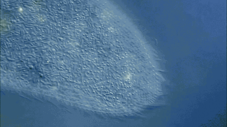
Paramecium, showing contractile vacuole and ciliary motion.Technique: Differential interference contrast, 350x-1000x. Shot by Mr. Ralph Grimm.Other specimens not pictured here include an "embryo of a black mastiff bat, a phantom midge larva, and a single-cell freshwater algae" according to Fast Company.For those hoping to catch a glimpse of some of these fascinating natural oddities, a selection of the winning photographs will be traveling through the U.S., Italy, Chile, Mexico, Brazil, Canada, Asia, and the Middle East in 2015.via Fast Co Create
ONE EMAIL. ONE STORY. EVERY WEEK. SIGN UP FOR THE VICE NEWSLETTER.
By signing up, you agree to the Terms of Use and Privacy Policy & to receive electronic communications from Vice Media Group, which may include marketing promotions, advertisements and sponsored content.

 Single-cell fresh water algae (desmids). Composite image including, concentric from the outside: Micrasterias rotata, Micrasterias sp., M. furcata, M. americana, 2x M. truncata, Euastrum sp. and Cosmarium sp. Technique: Confocal imaging, 400x. Shot by Dr. Igor Siwanowicz.
 Mouse embryonic fibroblasts showing actin filaments (red), mitochondria (green) and DNA (blue). Technique: Structured illumination microscopy (SIM) fluorescence, acquired with a 60x objective via Dr. Dylan Burnette.
 Paramecium, showing contractile vacuole and ciliary motion.Technique: Differential interference contrast, 350x-1000x. Shot by Mr. Ralph Grimm.Other specimens not pictured here include an "embryo of a black mastiff bat, a phantom midge larva, and a single-cell freshwater algae" according to Fast Company.For those hoping to catch a glimpse of some of these fascinating natural oddities, a selection of the winning photographs will be traveling through the U.S., Italy, Chile, Mexico, Brazil, Canada, Asia, and the Middle East in 2015.via Fast Co Create
